What to Expect From Breast Imaging Services at Radiology of Indiana
When it comes to breast health, waiting for answers can feel overwhelming. That’s why clarity, speed, and expertise matter.
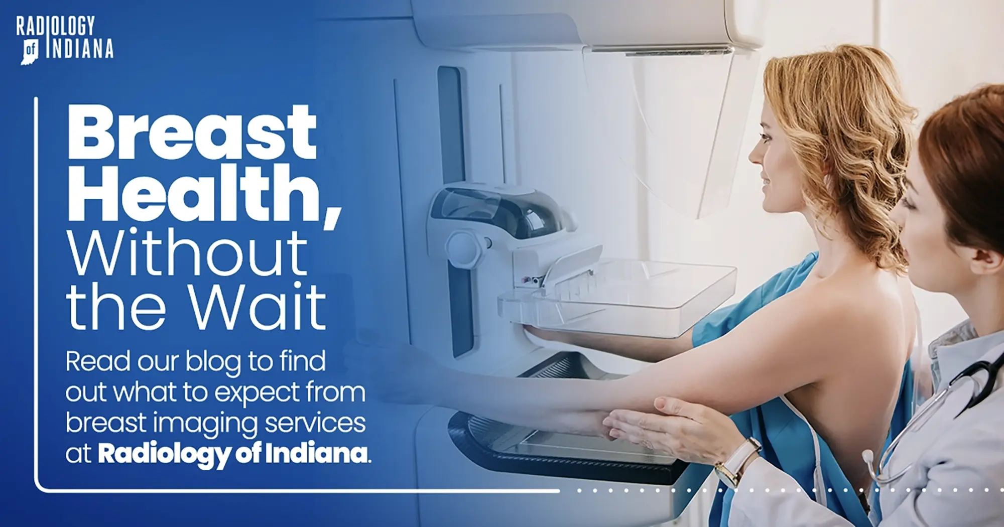
At Radiology of Indiana, our breast imaging services are built around one priority: delivering fast, accurate results in a supportive, patient-centered environment. From routine screenings to advanced diagnostic imaging, our team is focused on minimizing wait times and maximizing reassurance.
Comprehensive Breast Imaging, All in One Place
We offer a full range of breast imaging services, including:
- 3D Screening & Diagnostic Mammography
- Breast MRI
- Abbreviated Breast MRI (AB-MR)
- Ultrasound-Guided Breast Biopsy
- Stereotactic Breast Biopsy
Each exam is performed using state-of-the-art technology and interpreted by experienced breast radiologists who specialize in detailed, accurate analysis.
The Advantage of 3D Mammography
Digital breast tomosynthesis, also known as 3D mammography, is one of the most significant advances in breast cancer detection in decades.
Unlike traditional 2D mammography, 3D imaging allows radiologists to examine breast tissue layer by layer. This improves visibility and can:
- Detect invasive cancers earlier
- Reduce unnecessary callbacks
- Provide clearer evaluation of dense breast tissue
Our facility is accredited by the American College of Radiology, reflecting our commitment to high clinical standards and quality care.
Diagnostic Imaging When You Need It
If additional evaluation is required, we offer targeted diagnostic mammography, breast ultrasound, and breast MRI.
Breast MRI is especially valuable for women at higher risk, those recently diagnosed, or patients with dense breast tissue. We also offer Abbreviated Breast MRI (AB-MR), a shorter, lower-cost supplemental screening option for eligible patients.
Our process is designed to deliver timely results — with same-day interpretation whenever possible and reports sent to referring physicians within 24 hours.
Minimally Invasive Biopsy Options
If a biopsy is needed, we provide image-guided procedures that are precise, safe, and require little recovery time.
Both stereotactic and ultrasound-guided breast biopsies allow our radiologists to accurately target areas of concern while prioritizing patient comfort and minimizing scarring.
It’s important to remember: the majority of breast abnormalities are benign. If something unusual is detected, our team works quickly and thoughtfully to guide next steps.
A Patient-Centered Approach
We understand that breast imaging can bring anxiety. That’s why our entire process is built around:
- Rapid appointment scheduling
- Focused examinations
- Clear communication
- Compassionate care
From your first appointment to final results, your experience matters to us.
Schedule Your Appointment
Routine screening and timely evaluation are essential parts of breast health. To learn more or schedule your appointment, visit: https://www.radiologyofindiana.com/appointment/