Breast Cancer: The Importance of Imaging

Early detection can save lives; read our latest blog discussing breast cancer and the importance of imaging for detection and possibly treatment!

Breast Cancer Awareness

Join us this October as we recognize Breast Cancer Awareness Month because we aim to unite when cancer divides. Breast cancer is a form of cancer that develops in the breast when cells begin to multiply out of control. In the United States, one in eight women will develop breast cancer. Aside from skin cancer, breast cancer is the most common type of cancer among women. In 2022, the American Cancer Society estimates that approximately 43,250 women will die due to breast cancer, with a total of about 287,850 new invasive cases diagnosed.

The two most frequent forms of breast cancer are:

  • Invasive ductal carcinoma:Occurs when abnormal cells form within the milk ducts, then alter and attack breast tissue outside the ducts. Once this occurs, these cancer cells can spread to other body areas. The most common type of breast cancer, making up for a total of 80% of diagnoses.
  • Invasive lobular carcinoma – Beginning in the milk-producing glands known as the breast lobules, as the name invasive implies, this cancer can advance beyond the lobule. It can potentially reach one’s lymph nodes and other parts of the body. This form of breast cancer makes up around 10% of diagnoses.

Early Detection for Your Protection 

When discovered in its early stages, breast cancer has a survival rate of 99%, according to the National Breast Cancer Foundation. Three simple steps can help you remain proactive regarding breast cancer prevention. First, conduct a breast self-examination once a month at home. Familiarize yourself with how they feel and alert your doctor if changes arise. As the saying says, “feel for lumps, save your bumps.” The next step is a clinical breast exam; your physician or gynecologist completes a CBE at your annual examination. They are trained to notice any breast abnormalities or warning signs. The third and final step is a mammogram. This type of imaging allows a specialist to examine the breast tissue of targeted problem areas. Mammograms can detect breast lumps before they can be felt by hand.

Breast Cancer Awareness Pink ribbon.

Early detection is fundamental to treating breast cancer, with varied screening options readily available. Here are different types of radiological imaging used for breast cancer detection:

  • Breast MRI – A breast MRI (magnetic resonance imaging) is a diagnostic exam through a union of radio waves and powerful magnets that forms detailed images of the inside of the breast.
  • Breast Ultrasound – A screening test that utilizes sound waves to look within the breast. Breast ultrasounds also allow for specific breast changes to be monitored, such as a fluid-filled cyst that a mammogram may struggle to depict clearly.
  • Mammograms – Last but certainly not least is the most crucial screening test for breast cancer. Think of a mammogram as an X-ray of the breast, which can detect breast cancer as early as two years before a doctor can physically feel a tumor.

Breast cancer is an extremely difficult disease to experience or watch someone you love the experience. Therefore, raising awareness regarding means of prevention is essential moving forward. As actress and breast cancer survivor Ann Jillian once said, “There can be life after breast cancer. The prerequisite is early detection.”

Breast cancer research day - did you know 1 in 8 women will get breast cancer during her life_

Breast Cancer Awareness is more than just a month. Visit our website today to learn more about breast imaging and the various types provided!

Resources:

https://www.cdc.gov/cancer/dcpc/resources/features/breastcancerawareness/index.htm

https://www.cancer.org/cancer/breast-cancer/screening-tests-and-early-detection.html

https://www.hopkinsmedicine.org/health/conditions-and-diseases/breast-cancer/invasive-ductal-carcinoma-idc#:~:text=What%20is%20invasive%20ductal%20carcinoma,the%20walls%20of%20the%20duct


I changed the image to something a bit more modern. It’s a free image from Pexels!


I think this image was not used so I would find a new one to use from this month’s schedule!


Gotcha!!

What is Breast Density, and Why is it Important?

The American College of Radiology, the Society of Breast Imaging, and the National Comprehensive Cancer Network (NCCN), recommend that all women have yearly mammograms beginning at age 40. Women at high risk may benefit from starting earlier.

What is Breast Density?

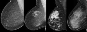
Breasts are made up of a mixture of fibrous and glandular tissue and fatty tissue. Your breasts are considered dense if you have a lot of fibrous or glandular tissue but not much fat. Density may decrease with age, but there is little, if any, change in most women.

  • 10% of women in the U.S. have almost entirely fatty breasts
  • 10% have extremely dense breasts
  • 80% are classified into one of two middle categories

How do I know if I have dense breasts?

Breast density is determined by the radiologist — the doctor who reads your mammogram. Your doctor should be able to tell you whether you have dense breasts based on where you fall on the density scale. Many states even have Breast Density Notification Laws, where doctors are required to inform their patients of their breast density following a screening Mammogram.

Why is Breast Density Important?

Having dense breast tissue may increase your risk of getting breast cancer. Dense breasts also make it more difficult for doctors to spot cancer on mammograms. Dense tissue appears the same on a mammogram as lumps, both benign and cancerous, making it difficult to distinguish lumps or differentiate between dense tissue and abnormalities. So, mammograms can be less accurate in women with dense breasts.

What are my screening options if I have dense breasts?

Even if you have dense breasts, you still need a yearly mammogram. A mammogram is the only medical imaging screening test proven to reduce breast cancer deaths. Many cancers are seen on mammograms even if you have dense breast tissue.

In breasts that are dense, cancer can be hard to see on a mammogram. Digital breast tomosynthesis (DBT), also called 3D mammography, provides images of the breast in “slices” from many different angles making some abnormalities easier to see. DBT increases the number of cancers seen without additional testing. Ultrasound (US) and magnetic resonance imaging (MRI) can help find cancers that can’t be seen on a mammogram. However, both MRI and US show more findings that are not cancer, which can result in added testing.

If you have dense breasts, please talk to your doctor. Together, you can decide which, if any, additional screening exams are right for you. If your breasts are not dense, other factors may still place you at increased risk for breast cancer — including a family history of the disease, previous chest radiation treatment for cancer and previous breast biopsies that show you are high risk.

Talk to your doctor and discuss your history. Even if you are at low risk, and have entirely fatty breasts, you should still get an annual mammogram starting at age 40.

Information sourced from the Mammography Saves Lives project, a collaboration between the Society of Breast Imaging and the American College of Radiology.

Breast Cancer Prevention

What Breast Cancer Screening Options are Available?

At Radiology of Indiana, our whole service is geared around the needs of women who want fast, expert and accurate analysis and immediate treatment where required. Our approach is highly empathetic towards individual patients. Your specific needs are important to us and will guide your treatment every step of the way, from the moment you come into one of our offices. We are proud to offer both 3D and 2D mammography, as well as Breast MRI at our various locations.

What is a 3D Mammogram?

Digital breast tomosynthesis, also known as 3D mammography, is a revolutionary new screening and diagnostic breast imaging tool that has improved the early detection of breast cancer. During the 3D part of the exam, an x-ray arm takes multiple images in seconds as it sweeps over the breast. Images are displayed as a series of thin sliced layers that can be viewed by radiologists as individual images or in an interactive animation. 3D mammography is used in combination with 2D digital mammography.

What are the benefits of 3D Mammography? 

A 3D mammogram is very similar to having a 2D conventional mammogram; however it is clinically proven to be more accurate. This type of mammogram allows doctors to examine your breast tissue layer by layer so that fine details are more visible and not hidden by the tissue above or below. 3D mammography is known as the biggest breakthrough in breast cancer detection in the last 30 years.

What is Breast MRI?

MRI is a sophisticated technology that uses a magnetic field to provide three dimensional images of the breast. Mammography remains the test of choice in screening for breast cancer, though in certain very high risk women, MRI may be helpful. MRI doesn’t replace mammography – it’s a different imaging technique that provides additional information for the detection and characterization of breast disease.

Who Should Use Breast MRI?

  • Women recently diagnosed with breast cancer.
  • Women at especially high risk for breast cancer, including:
    • Those with altered BRCA1 and BRCA2 genes.
    • First degree relative of altered BRCA1 and BRCA2 genes.
    • Women with increased risk of breast cancer based on relatives diagnosed with breast cancer, history of ovarian cancer, and other factors.
  • Women treated for breast cancer, to evaluate response to treatment.
  • Women concerned about rupture of a silicone implant.

Where Can I Schedule Preventative Breast Screenings?

You can visit this page to schedule a 3D mammogram or a Breast MRI consultation.